Osteogenesis imperfecta is a group of genetic disorders marked by extremely fragile bones that break or fracture easily. Often from little or no apparent trauma or cause.
The condition is also called brittle bone disease. The severity of osteogenesis imperfecta varies from person to person, even among individuals of the same family.
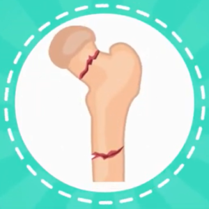
It ranges from mild to severe. A person may have just a few or as many as several hundred fractures in their lifetime.
Most cases are mild resulting in few bone fractures, the severe form can cause hearing loss, spinal cord problems, and heart failure as well as permanent deformities.
- The condition can be life-threatening if it occurs in babies either before or after birth.
- The condition affects both males and females equally and affects about 1 in 15,000 people.
Causes:
Osteogenesis imperfecta is a genetic disorder that is caused by a mutation in the COL1A1 or COLIA2 gene inherited in an autosomal dominant pattern or occurs via a new mutation.
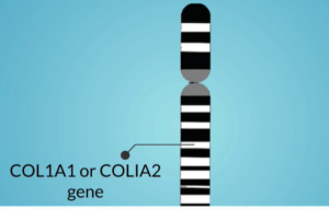
This means that a child inherits the defective gene from one of their parents. Or neither parent has it but the defect occurs due to a spontaneous mutation (change) in the gene, and it stops working properly. This gene produces a protein (type 1 collagen), a major component of the connective tissues in bones.
Type 1 collagen is also important in forming teeth, ligaments, and sclera (the white outer tissue of the eyeballs)
Types:
There are eight types with type 1 being the least severe and type 8 being the most severe.
The first four types are the most common while the last four are extremely rare, and most are subtypes of type 4 osteogenesis imperfecta (OI),
Type 1 (OI):
This is the mildest and the most common of the condition. In this type, the body produces the normal quality of collagen but in insufficient quantities.
These results in mildly fragile bones a bone fracture may occur due to mild trauma. The bone fracture usually occurs during childhood through puberty and maybe less common after puberty.
Features may include:
- Bluish discoloration of the sclera (blue sclera)
- Abnormalities in the middle or inner ears resulting in hearing impairment
- Loose joints
- Low muscle tone
- Bone fractures easily
- Abnormal outward curvature of the upper spine (Scoliosis)
- An abnormal outward curvature of the upper spine (Kyphosis)
- Slight protrusion of the eye
Type 2 (OI):
This type is the most severe type and can cause life-threatening complications at, or shortly after birth.
In this type, the body produces insufficient collagen or produces collagen that is of poor quality. Most children born with type 2 die within the first year of life due to respiratory failure, due to underdeveloped lungs.
This type is sub-classified into groups A, B, C which are distinguished by bone formation seen only on x-ray.
Features:
- Underdeveloped lungs
- Severe bone deformity and small stature
- Narrowed chest
- Type 2A have broad and short long bones with broad and beaded ribs
- Type 2B demonstrates broad and short long bones with thin ribs with little or no beading
- Type 2C shows thin and longer long bones with thin and beaded ribs
Type 3 (OI):
In type 3, enough collagen is made but it is defective and causes the bone to break easily. Bone deformities are common and present at birth. They may become worse as affected children age.
Features:
- Severe bone deformities
- Loose joints
- Triangular face
- Poor muscle tone in arms and legs
- Early loss of hearing is possible
- Discoloration of the sclera
- Short Stature
- Respiratory problem is possible
- Brittle, discolored teeth may also be present
Type 4 (OI):
This type is the most variable because symptoms range from mild to severe. In this type, enough collagen is made but it is not of high quality.
Features:
- Bone fracture easily, especially before puberty
- Short stature
- Mild to moderate bone malformation
- Hearing impairment
- Spinal curvature
- Bowed legs that may lessen with age.
Diagnosis:
The condition is diagnosed by taking x-rays. This allows the doctor to see current and broken bones as well as view defects in the bones.
A laboratory test may be done. This may include taking blood or tissue samples for genetic testing.
Treatment:
No cure exists for this condition. But, there are supportive therapies that help reduce the risk of broken bones and may increase the quality of life.
- Physiotherapy is used to strengthen muscles.
- Physical aids such as crutches, wheelchairs, grabbing arms
- Medicine to reduce any pain
- Bisphosphonate medications to strengthen a child’s bone
- Low-impact exercise for bone building
- Surgery may include inserting a metal rod in the long bones to improve strength
For more information talk to a healthcare provider.
If you have any questions about Osteogenesis Imperfecta, please feel free and leave a comment.
Do share this blog with your friends and family!