Overview:
A joint is defined as a connection between two bones in the skeleton. There are many ways in which joints can be classified.
They can be classified according to their structure, their mobility, and their range of motion.
Joint classification:
Structure
There are three distinct types-
- Synovial joint
- fibrous joint
- Cartilaginous joints
Synovial joint:
The synovial joint as it’s the most common joint found in the human body. Synovial joints have several characteristic features as a sagittal illustration of the knee joint.
First of all, synovial joints are surrounded by an articular capsule. This capsule consists of an outer fibrous layer that helps us to stabilize the joint and an inner synovial layer that absorbs and secretes synovial fluid lubricating the joints.
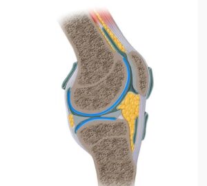
The articular surfaces of a synovial joint are covered in hyaline cartilage. This cartilage is also known as articular cartilage and acts to reduce friction and assists in shock absorption.
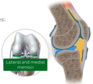
Additional structures may also be found within synovial joints such as articular discs.
We can see examples of these in the knee joint- the medial and lateral menisci.
The bursae, which are small sacs lined by the synovial membrane and filled with synovial fluid.
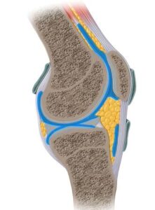
The bursae act to reduce friction caused by muscles and tendons which are located over bony joints.
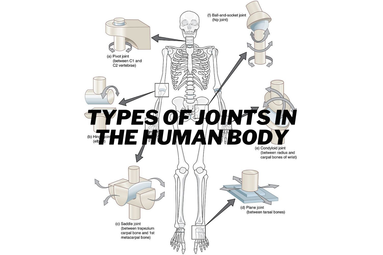
The synovial joints can be further sub-classified according to the shape of their articular surfaces and their range of motion.
These include,
- Ball and socket joint
- Hinge joint
- Pivot joint
- Condylar joint
- Saddle joint
- Plane joint
Fibrous joint:
In a fibrous joint, the bones are bound by a tough fibrous connective tissue. These joints exhibit little to no mobility.

Types of fibrous joints:
- Sutures
- Gomphoses
- Syndesmoses
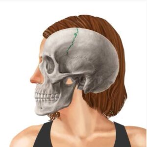
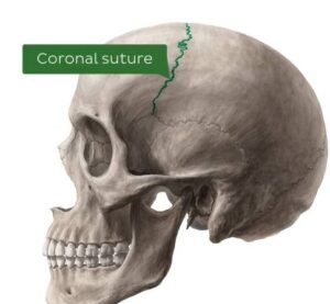
The sutures, which are fibrous joints found exclusively between the bones of the skull.
This is the coronal suture. The suture connects the frontal bone to the parietal bones via suture ligaments.
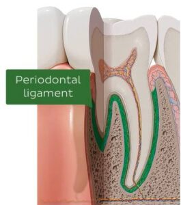
Gomphosis is found in the mouth, where the roots of the teeth articulate with the dental alveoli at the dentoalveolar joints.
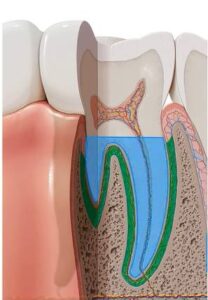
The tooth is bound into its socket by the strong periodontal ligament.
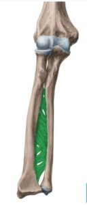
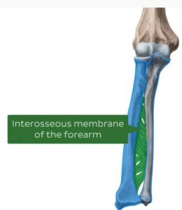
The syndesmosis, which is formed by ligaments and a strong membrane that holds two bones in place. The interosseous membrane runs between the radius and ulna.
Cartilaginous joint:
In a cartilaginous joint, the bones are connected by fibrocartilage or hyaline cartilage.
These joints can be subclassified into two types
- Synchondroses
- Symphyses
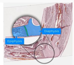
Synchondroses or primary cartilaginous joint, where the bones are connected by hyaline cartilage.
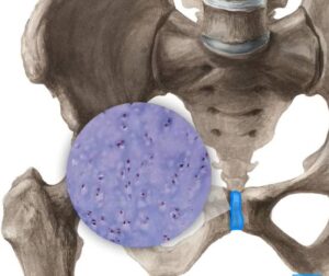
This type of joint is found between the diaphysis and epiphysis of a growing long bone and will eventually become completely ossified in adulthood.
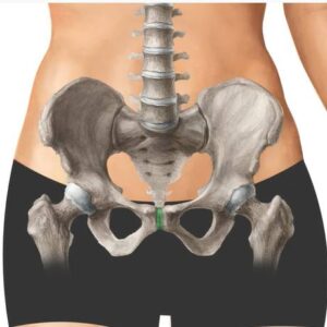
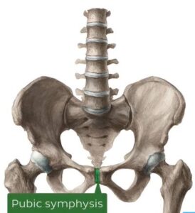
The second type of cartilaginous joint is known as a symphyses or secondary cartilaginous joint, where the bones are connected by fibrocartilage.
This type of joint is found primarily along the midline of the body; for example, the pubic symphysis.
Mobility:
Diarthrosis:
A diarthrosis is a freely mobile joint, and an example of this is the knee joint which is a synovial hinge joint. It’s worth noting that every synovial joint is a diarthrosis.
Amphiarthrosis:
An amphiarthrosis is a slightly mobile joint, and an example of this is the pubic symphysis. Which is a secondary cartilaginous joint.
The syndesmosis, which is a fibrous joint, is also an example of amphiarthrosis.
Synarthrosis:
A synarthrosis, which is an immobile joint, and an example of this is the coronal suture of the skull.
Other examples, of this type of joint include,
- Gomphosis
- Synchondrosis, which is a primary cartilaginous joint.
Range of Motion:
When classifying a joint based on its range of motion, it’s important to first understand the various axes of movement that the movements are occurring along in joint movement, there are three main axes
Axes of Movement:
There are three main axes,
- The sagittal axis, which passes horizontally from anterior to posterior.
- The frontal axis, which passes horizontally from left to right.
- The vertical axis, which passes vertically from superior to inferior.
Types:
- The joint that can move back and forth along a single axis is called uniaxial. Examples of these are the hinge joint and the pivot joint.
- Joints that move about two distinct axes are called biaxial. For example, the condylar joint and the saddle joint.
- Joints that can move through all three axes are called polyaxial or multiaxial. The only example of this is the ball and socket joint.
Synovial Joints: Anatomy
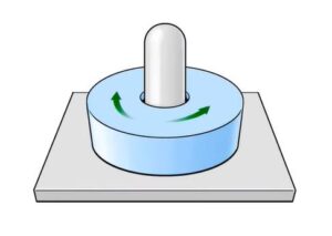
Ball and socket joint:
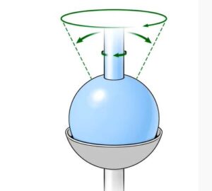
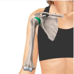
The ball and socket joint, which is also known as the spheroid joint. This is the only polyaxial joint and therefore, the most mobile of all joint types.
The movements that occur at these joints are,
- Flexion and extension, which occur around a frontal axis.
- Adduction and abduction, which occur around a sagittal axis.
- Internal rotation and external rotation, which occur around a vertical axis.
Two examples of this joint are the acetabulofemoral joint or the hip joint and the glenohumeral joint, which is known as the shoulder joint.
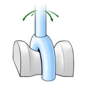
Hinge joint:
The hinge joint, which is only one axis of motion making it uniaxial.
The movements that occur at these joints are,
- Flexion
- Extension
Two examples of these joints are the tibiofemoral joint or the knee joint and the elbow joint.
So, we use our hinge joints when we show off to flex our biceps!
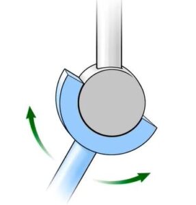
Pivot joint:
Like the hinge joint, the pivot joint is also uniaxial. So, pivot joints, also known as rotary joints, allow movement around a single axis –this movement being rotation.
Condylar joint:
The condylar joint, which is also known as the ellipsoid joint. Condylar joints allow movement around two axes that are at right angles to each other. Therefore, they are described as biaxial joints.
An example of this joint is the radiocarpal joint or the wrist joint.
The movements that take place at this joint are,
- Radial deviation and ulnar deviation, which occur around the sagittal axis.
- Flexion and extension, which occur around the frontal axis.
- These movements combine to produce circumduction of the wrist joint.
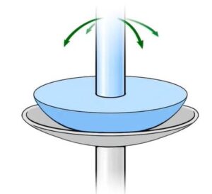
Saddle joint:
Similar to the condylar joint, the saddle joint is a biaxial joint.
The movement that takes place at this joint is,
- Abduction and Adduction
- Flexion and Extension
- Circumduction
An example of such a joint is the Carpometacarpal joint of the thumb.
Plane joint:
The plane joint is also known as the gliding joint. The plane joint performs a sliding or a gliding movement, where one bone moves across the surface of another.
An example of this joint is the acromioclavicular joint, which increases the flexibility of the shoulder.
For more information talk to a healthcare provider.
If you have any questions about Types of joints in Humans, please feel free and leave a comment.
Do share this blog with your friends and family!