Muscles of the hip:
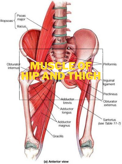
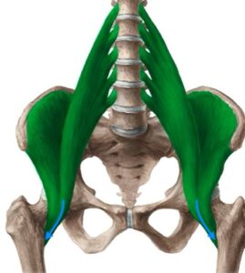
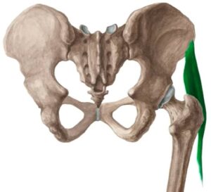
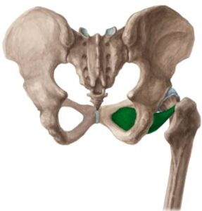
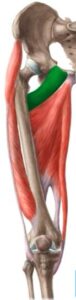
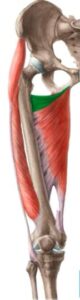
Anterior hip muscles: Large one you can see from an anterior perspective and highlight in green, which is the psoas major muscle. The psoas major originates from the bodies of the vertebrate T12 to L4 and the costal processes of the vertebrae L1 to L5.
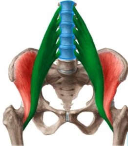
Located a bit more laterally, we find another muscle which is known as the iliacus muscle.
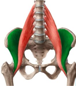
The iliacus muscle originates from the iliac fossa. The psoas major and the iliacus are usually distinguished as one muscle, and this is known as the iliopsoas muscle.
These muscles have different points of origin, however, they come together to pass underneath the inguinal ligament and into the region of the thigh to insert onto the lesser trochanter of the femur.
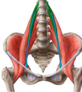
The last muscle of the anterior hip, we are going to be talking about is the psoas minor muscle. This is a small muscle that runs along the surface of the psoas major. This muscle is sometimes not mentioned as it is often absent. Around about 40%-70% of people do not have this muscle.
Anterior Hip Muscles: Function/Innervation/Blood Supply
- Function: The iliopsoas is the most powerful flexor of the thigh at the hip joint.
- Innervation: The lumbar plexus innervates the psoas major and psoas minor muscles.
- Blood Supply: The iliopsoas muscle receives its blood supply from the iliolumbar artery and the medial femoral circumflex artery.
Muscles of the hip: Superficial gluteal muscles
There are four superficial gluteal muscles,
1. Gluteus maximus muscle:
It’s the most famous muscle that defines the buttocks. The gluteus maximus originates from the surface of the ilium posterior to the posterior gluteal line and the posterior inferior surface of the sacrum and the coccyx.
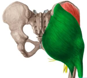
It inserts onto the gluteal tuberosity of the femur and the iliotibial tract.
2. Gluteus medius muscle:
The gluteus medius originates from the gluteal surface of the ilium and inserts onto the greater trochanter.

3. Gluteus minimus muscle:
Deep to the gluteus medium muscle, we find the gluteus minimus. The gluteus minimus also originates from the gluteal surface of the ilium. Inserts onto the greater trochanter of the femur.
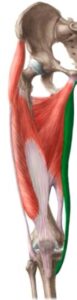
4. Superficial gluteal muscle:
The muscle extends from its origin at the anterior superior iliac line to its insertion at the iliotibial tract.
Muscles of the hip: Superficial gluteal muscles (Function/Blood Supply)
Function: Extension, Abduction, Rotation of the thigh at the hip joint, Stabilize the pelvis.
Blood supply: The muscles receive their blood supply from the superior gluteal artery and the inferior gluteal artery.
Muscles of the Hip Deep Gluteal:
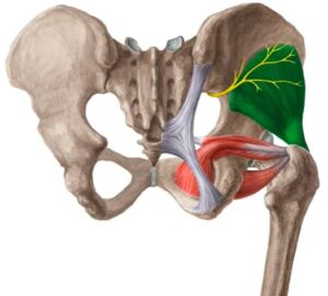
Piriformis muscle: The muscle originates from the pelvic surface of the sacrum and is insert onto the greater trochanter of the femur.
Obturator internus muscle: The obturator internus originates from the obturator membrane and inserts onto the greater trochanter and trochanteric fossa.
Superior gemellus muscle: This muscle originates from the ischial spine and inserts onto the greater trochanter of the femur.
Inferior gemellus muscle: The inferior gemellus originates from the tuberosity of the ischium and inserts onto the greater trochanter.
Quadratus femoris muscle: This muscle originates from the tuberosity of the ischium and inserts into the intertrochanteric crest.
Muscles of the Hip Deep Gluteal: (Function/Innervation/Blood Supply)
Functions: lateral rotation of the thigh at the hip joint.
Innervation: Is supplied by the sacral plexus.
Blood supply: The muscle receive their blood supply from the superior gluteal artery and the inferior gluteal artery.
Muscles of the Thigh: Anterior Compartments
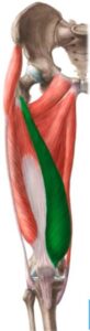
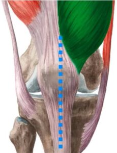
Sartorius muscle: This is the longest muscle in the human body and it extends from its origin at the anterior superior iliac spine. All the way to its insertion on the medial surface of the tibia.
This muscle has various functions including flexion of the thigh and knee, lateral rotation of the thigh, and medial rotation of the knee.
Quadriceps femoris muscle: The muscle is formed by four muscles –the rectus femoris, vastus lateralis, vastus intermedius, and vastus medialis.
These muscles all have different sites of origin. However, they all insert into the quadriceps tendon.
Let’s take a look at these muscles individually, located most anteriorly, we have the rectus femoris muscle, and this muscle originates from the anterior inferior iliac spine and the supraacetabular sulcus.
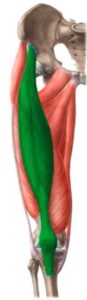
The vastus lateralis muscle is located laterally and it originated from the linea aspera femoris and the greater trochanter.
In the middle, we can see another vastus which is known as the vastus intermedius muscle, and this muscle originates from the shaft of the femur.
The next muscle is found medially and is known as the vastus medially muscle. This muscle originates from the linea aspera femoris and the intertrochanteric line.
The last muscle of the anterior compartment is the articularis genu muscle, and it lies deep to the vastus intermedius
This small flat muscle originates from the anterior distal femoral shaft and inserts onto the knee joint capsule.
Muscles of the anterior compartment:
Function:
- Extension of the leg at the knee joint.
- Flexion the thigh at the hip joint.
Innervation:
These muscles receive their innervation from the femoral nerve and their blood supply from the femoral artery and the deep femoral artery.
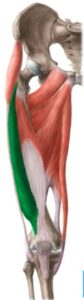
Muscles of the thigh: Medial compartment
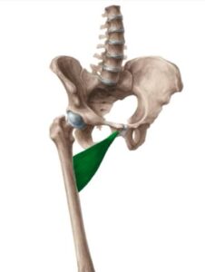
Obturator externus muscle: This muscle originates from the obturator foramen and the obturator membrane and inserts at the trochanteric fossa.
The pectineus muscle originates from the iliopubic eminence and the pectineal line of the pubic bone and inserts at the linea asper femoris and the pectineal line of the femur.
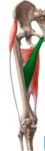
Located most medially, the gracilis muscle is an exception within this group of the thigh muscles because it inserts on the tibia. The muscle originals from the inferior pubic ramus insert on the proximal medial surface of the tibia.
Adductors of the Thigh:
Adductor Brevis muscle: The word Brevis means ‘short’ in Latin and adductor brevis is quite a short muscle. This muscle originates from the inferior pubic ramus and inserts on the linea aspera femoris.
Adductor longus muscle: The adductor longus originates from the pubic symphysis and the superior pubic ramus and inserts on the linea aspera femoris.
Adductor Magnus muscle: This muscle originates from the inferior pubic ramus, ramus of the ischium, and tuberosity of the ischium. It inserts on the linea aspera femoris and the adductor tubercle.
Adductor minimus muscle: The adductor minimus originates from the inferior pubic ramus and inserts on the linea aspera femoris.
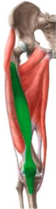
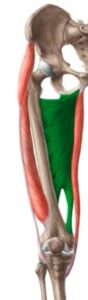
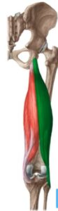
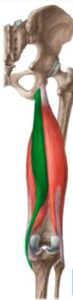
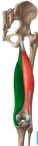
Muscles of the Thigh: Posterior compartment
The muscles of the posterior compartment are also known as the hamstring muscles.
Biceps femoris muscle: It originates from the Sacrotuberous ligament, linea Aspera femoris, and tuberosity of the ischium. It then inserts onto the head of the fibula.
Semitendinosus muscle: This muscle is located medially, and muscle originates from the sacrotuberous ligament and tuberosity of the ischium and it inserts on the proximal tibia medial to the tibial tuberosity.
Semimembranosus muscle: This muscle originates from the tuberosity of the ischium and inserts on the medial condyle of the tibia and the oblique popliteal ligament.
For further advice do reach out to your local doctor or family doctor.
Do share this blog with your friends and family!